
Tests & Procedures
Imaging Tests & Scans

There are a variety of imaging techniques that doctors use to understand what’s happening inside your body. For gastroenterology and digestive health, the focus is mainly on the abdomen. Scans and imaging tests can look at different organs inside the abdomen including the small intestine, liver, bile ducts, gall bladder, pancreas, spleen, kidneys, and uterus.

Ultrasound scan.
An ultrasound scan is a safe, simple, and quick test that is often ordered to look for causes of abdominal pain or bloating. It (usually) gives good views of the gallbladder and bile ducts, the kidneys, the uterus, and the ovaries. It is less useful for looking at the stomach and the intestines, and sometimes the pancreas can be hidden by gas in the bowel. A special type of ultrasound scan (liver elastography or ‘Fibroscan’) can measure the amount of fat and scar tissue in the liver.
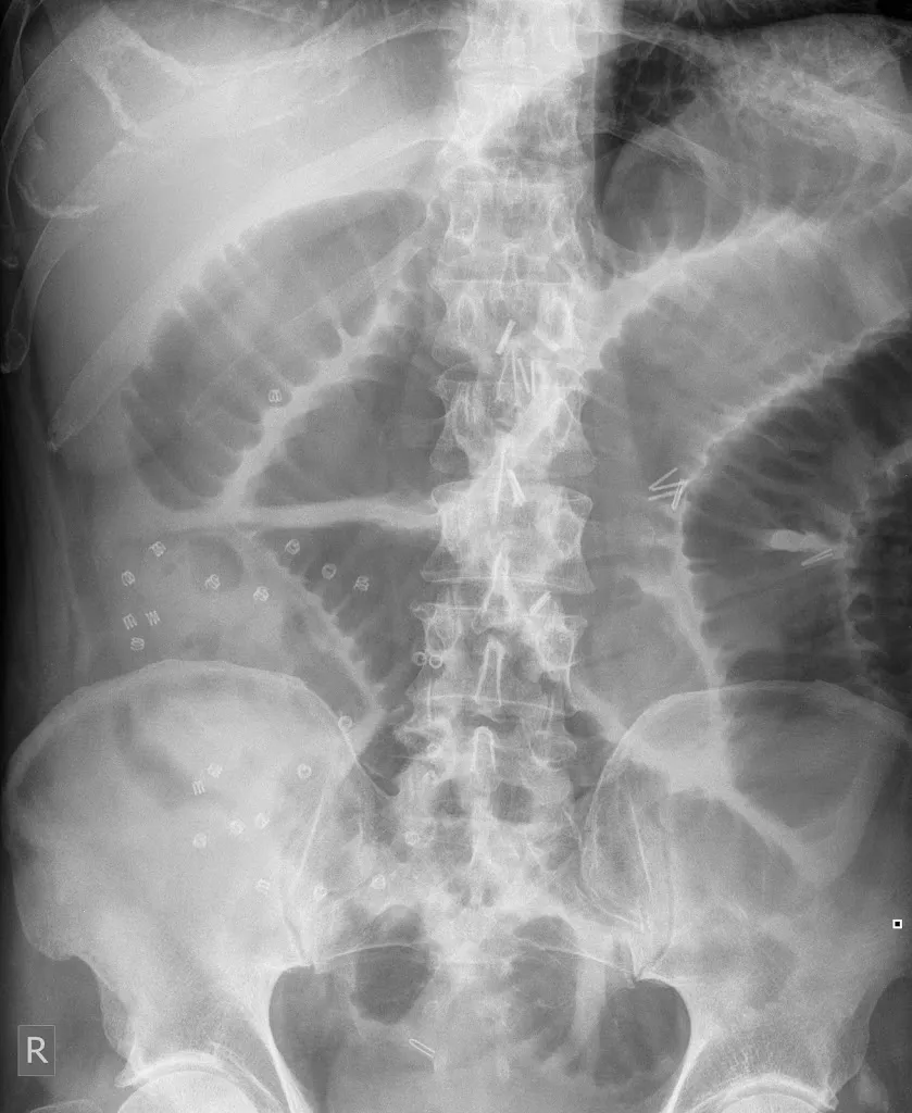
Abdominal X-ray
Your bowels do not show up very well on ordinary X-rays, but it is possible to get an idea of the amount of gas and stool in the intestines – particularly the colon – on a plain X-ray of the abdomen. X-rays can be useful to investigate constipation and bloating, or conditions where there is a lot of inflammation in the bowel such as Ulcerative Colitis.

CT scan
A CT (computed tomography) scan uses X-rays and computers to generate pictures of cross-sectional ‘slices’ through the body. During a CT scan, you will lie flat on a couch which slides inside a giant ring (or doughnut). A CT scan is not at all claustrophobic and typically lasts 5-15 minutes. Just before the scan, you may be given an injection of a CT contrast dye. The contrast dye is not dangerous unless you have kidney problems or certain allergies. A CT can give very clear pictures of the solid organs in the abdomen such as the pancreas, liver, and kidneys. It is also a very good test to spot significant inflammation in the wall of the bowel (e.g. diverticulitis, severe colitis), appendix (e.g. appendicitis), and gallbladder. CT is less good at looking at the stomach or problems inside the intestines or bile ducts but can normally show if there is a problem severe enough to cause a blockage.
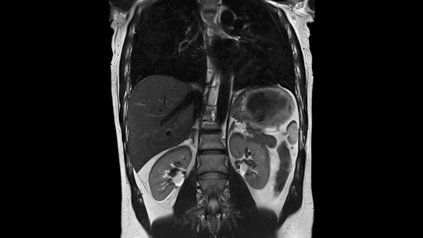
MRI scan
An MRI (magnetic resonance imaging) scanner uses powerful magnets to generate pictures of the inside of the abdomen. During an MRI scan, you will lie flat on a couch which slides inside a tunnel. The scan usually takes 25-45 minutes. An MRI scan is a very safe test (although it is rather noisy) and does not expose you to X-rays. However, because you are lying in quite a narrow tunnel, an MRI scan can feel claustrophobic, and for some people, this can make an MRI scan very difficult to tolerate. If necessary you can be referred to a hospital with an ‘open’ MRI scanner. In an ‘open’ MRI scan there is no tunnel and so claustrophobic patients find it a lot easier to tolerate. Because an MRI scan uses powerful magnets, you should not have an MRI scan if you have a pacemaker, implantable cardiac defibrillator, or have had a shrapnel injury. Metal joint replacements are ok. An MRI scan can give very clear images of the gallbladder and bile ducts, the pancreas, the liver, and the small bowel.
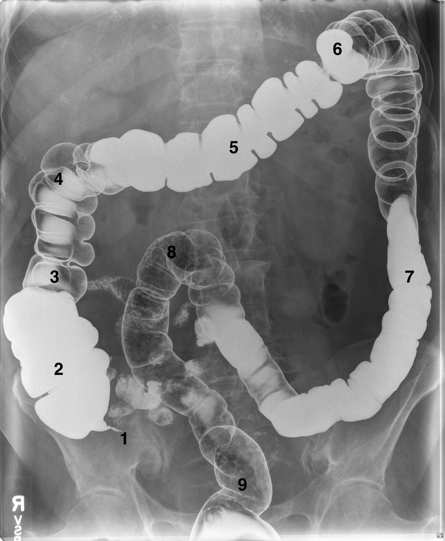
Barium Swallow and Barium Meal
The esophagus and stomach do not show up very well on X-rays and most other imaging tests. Barium can be used to highlight these organs as X-Rays do not pass through barium which can act as an X-Ray ‘dye’. If you drink a white solution containing barium sulfate, the barium will coat the lining of the esophagus and stomach and so their outline shows up clearly on X-Rays. This can be used to show up abnormalities in the lining or the anatomy of the esophagus and stomach such as ulcers, narrowed areas, or cancers. This was the main use for Barium tests in the days before endoscopy – however, these days gastroscopy is a much more useful test for looking at the lining of the gut. However, by taking several X-Rays in quick succession, a video of how barium passes through the esophagus and stomach can be obtained. This gives very useful information on the motility and functioning of the esophagus and stomach – e.g. how well the muscles in the esophagus and stomach are contracting, whether they are emptying properly, and whether stomach contents can easily reflux back up into the esophagus

For professional Imaging Tests and scans, related advice and queries feel free to get in touch with Dr Neil